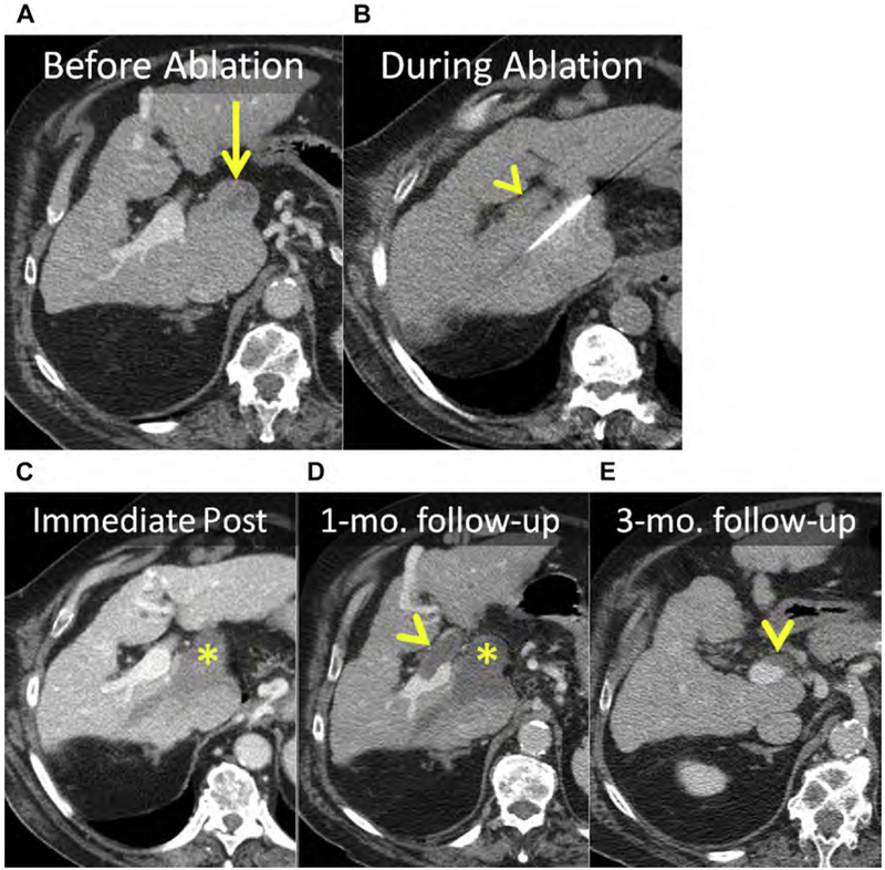
Fig. 5.
(A) Enhanced CT shows an HCC in the caudate (arrow). (B) Unenhanced CT after placement of MW antenna. Note the proximity of the main portal vein (arrowhead). (C–E) Serial follow-up enhanced CT immediately (C), 1 month (D), and 3 months after MW ablation showing the ablation (asterisks) without LTP. (C) The main portal vein was patent immediately after ablation. (D) At 1-month follow-up, there was thrombosis of the main and right portal veins (arrowhead) that (E) organized and partially resolved after anticoagulation therapy (arrowhead).

