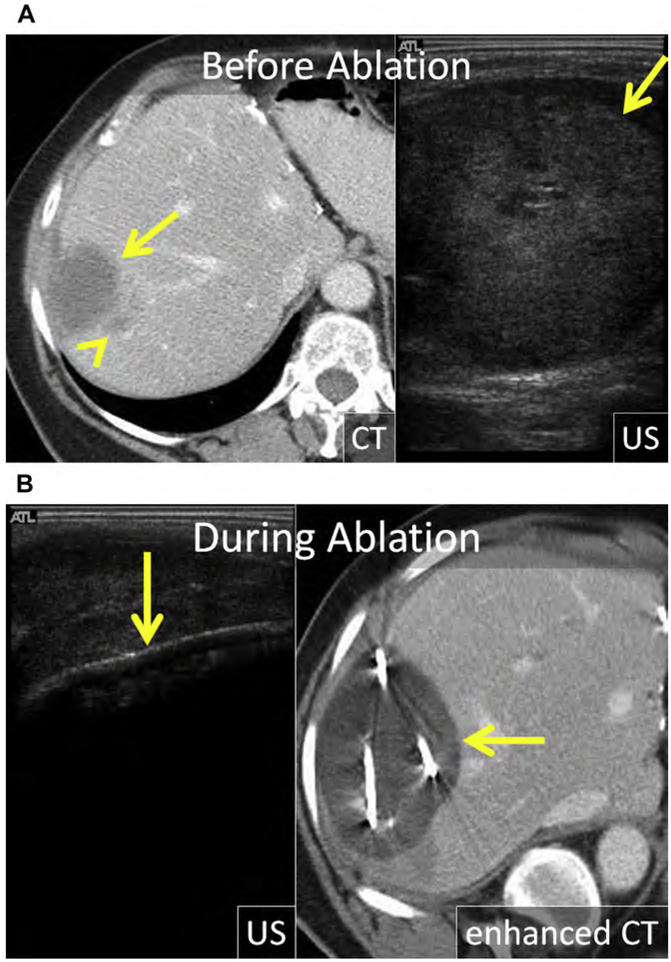
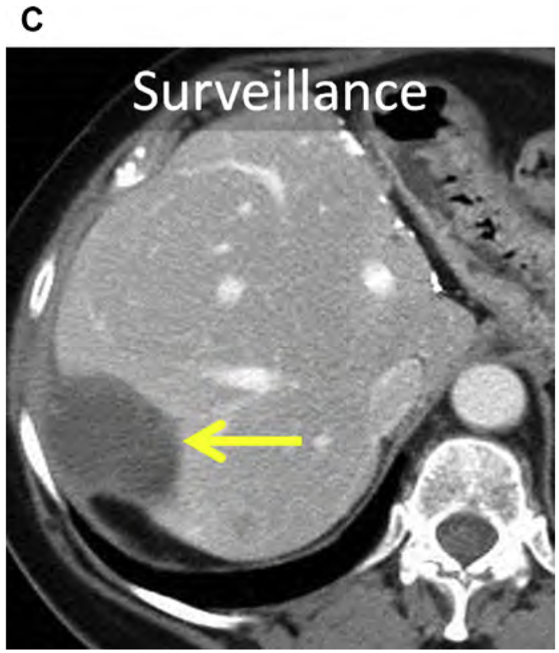
Fig. 9.
(A) CRLM in the right liver (arrow) with a dominant satellite tumor along the inferomedial margin (arrowhead). The mass is heterogeneous and echogenic at US. (B) US and enhanced CT during cryoablation. The ice ball is visible at US and CT (arrow); however, shadowing at US precludes evaluation of the deep margin. The distance between the visible ice and the lethal isotherm within the ice ball depends on local factors, including perfusion-mediated tissue warming. (C) Enhanced CT 1 year after cryoablation; there is no evidence of LTP (arrow).


