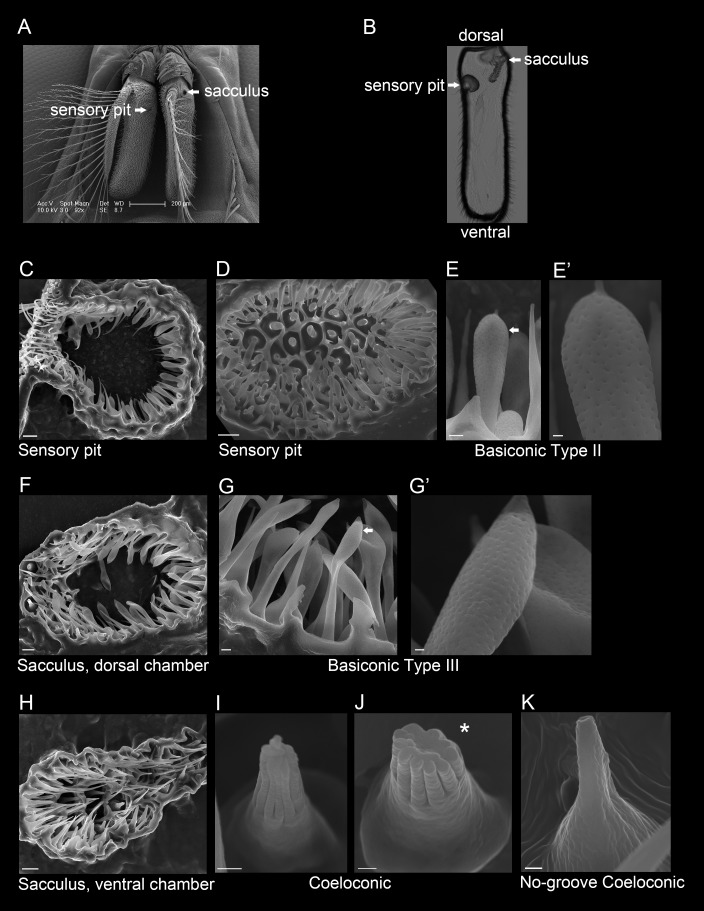
Fig 2. The sensory pit and sacculus of the G. morsitans antenna.
(A) Scanning electron micrograph of a G. morsitans head with arrows indicating the openings of the sensory pit and sacculus. (B) Transmitted light image of an antennal cross section (coronal plane) showing the sensory pit and sacculus that open to the medial and lateral sides of the antenna, respectively. (C) Micrograph of a sectioned sensory pit with its opening to the antennal surface at the upper left. (D) Micrograph of the sensory pit, showing a top-down view of the olfactory sensilla that line the pit. (E, E’) The sensory pit is lined with basiconic Type II sensilla. The arrow in E indicates the sensillum shown in E’. (F) Dorsal chamber of the sacculus (medial is left, lateral is right) that is lined with basiconic Type III sensilla (G, G’). The arrow in G indicates the sensillum shown in G’. (H) Ventral chamber of the sacculus (medial is at left, lateral is at right) that is lined with coeloconic (I, J) and no-groove coeloconic sensilla (K). The asterisk in panel J marks an axial view of a coeloconic sensillum that had its tip cut off during cryosectioning, fortuitously revealing its inner structure. Scale bars = 200 μm for A; 5 μm for C, D, F and H; 1 μm for E, G; and 0.25 μm for E’, G’, I, J, and K.

