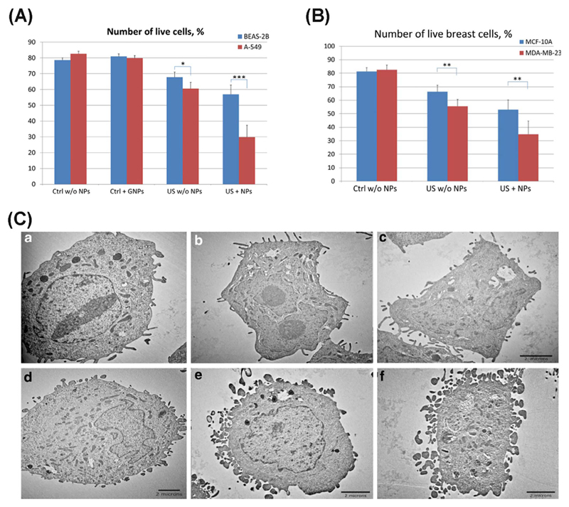
Fig. 3.
(A) BEAS-2B normal lung cells and A549 lung cancer cells after US treatment with gold NPs and the corresponding control cells (untreated with or without US or NPs). (B) MCF-10A normal breast cells and MDA-MB-231 breast cancer cells with/without US treatment and super-paramagnetic iron oxide NPs and the corresponding control (untreated) cells *represents P < .05, **represents P < .01, ***represents P < .001. (C) TEM images of H-184B5F5/M10 normal breast cells (a–c) and MDA-MB-231 breast cancer cells (d–f) cells, where a and d are control samples; b and e correspond to cells treated with US; c and f show the cells after combined treatment with US and magnetic NPs [105].

