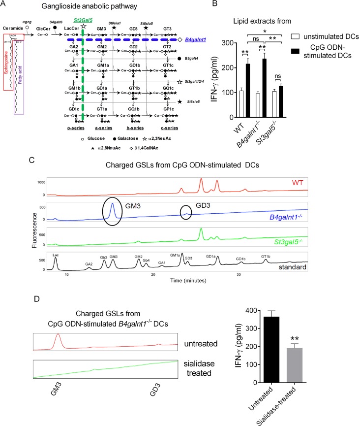
Fig 1. Endogenous ligand(s) for iNKT cells in CpG ODN-stimulated DC is/are simple sialylated gangliosides.
(A) Ganglioside anabolic pathway including species and key enzymes involved is depicted. (B) Base-treated charged lipids extracted from unstimulated or CpG ODN-stimulated DCs derived from WT, B4galnt1−/−, or St3gal5−/− mice were exposed to DCs (1/500 in DMSO; 100,000 DCs) for 16 h and, after extensive washes, were cocultured with iNKT cell-enriched liver MNCs (500,000 cells) for 24 h. In this setting, IFNγ secretion is measured as a read-out for iNKT cell activity, although we cannot exclude that it can also be produced by other innate lymphocytes (e.g., NK cells) through bystander effect. Data represent the mean ± SEM of three independent experiments performed in duplicates. (C) Glycosphingolipids were extracted and isolated from CpG ODN-stimulated B4galnt1−/− and St3gal5−/− DCs, as described in the methods. Charged species were then separated from neutral species using DEAE ion-exchange chromatography prior to NP-HPLC. (D) Sialidase A-treated or untreated base-treated charged lipids extracted from CpG ODN-stimulated DCs were tested for iNKT cell antigenicity as in Fig 1B. Data represent the mean ± SEM of two independent experiments performed in triplicates. **P < 0.01; *P < 0.05. Underlying data used in the generation of this figure can be found in S1 Data. CpG, cytosine-phosphate-guanine; DC, dendritic cell; DEAE, diethylaminoethyl; MNC, mononuclear cell; IFNγ, interferon gamma; iNKT, invariant natural killer T; NK, natural killer; NP-HPLC, normal-phase high-pressure liquid chromatography; ODN, oligodinucleotide; WT, wild-type.

