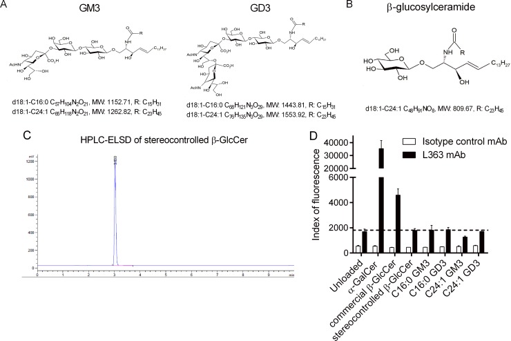
Fig 3. Structure and quality control of ganglioside species.
(A) Structures and formulas of synthetic GM3 and GD3 gangliosides. (B) Structure and formula of synthetic β-glucosylceramide (C24:1). (C) HPLC-ELSD of stereo-controlled β-GlcCer. (D) Plate-bound CD1d dimers loaded with various glycolipids were incubated with the PE-conjugated L363 mAb. Fluorescence at 575 nm was measured using a microplate reader. One representative experiment out of two performed in triplicates is shown. Underlying data used in the generation of this figure can be found in S1 Data. β-GlcCer, β-glucosylceramide; CD1d, cluster of differentiation 1d; HPLC-ELSD, high-performance liquid chromatography-evaporative light scattering detector; mAb, monoclonal antibody; PE, phycoerythrin.

