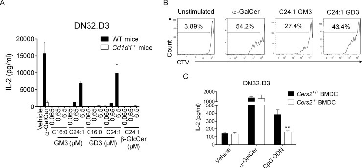
Fig 5. In vitro activity of gangliosides on iNKT cells.
(A) DCs derived from WT or CD1d1−/− mice were cultured with the mouse iNKT hybridoma DN32.D3 in presence of purified glycolipids at the indicated concentrations or vehicle in complete RPMI media for 24 h. IL-2 production was quantified by ELISA. Means ± SEM of one representative experiment out of three performed in triplicates are shown. (B) Cell Trace Violet-labeled spleen cells from C57BL/6 mice were cultured in absence or presence of α-GalCer (100 ng/ml), C24:1 GM3 (6.5 μM), or GD3 (6.5 μM) in complete RPMI plus recombinant IL-2. After 72 h, cells were harvested and analyzed by flow cytometry for CTV detection in live iNKT cells. Dot plots are representative of one experiment out of three. (C) DCs derived from Cers2+/+ or Cers2−/− mice were cultured with DN32.D3 in presence of α-GalCer (20 ng/ml), CpG ODN (2 μg/ml), or vehicle in complete RPMI media for 24 h. Means ± SEM of IL-2 concentrations of one representative out of three experiments performed in duplicates are shown. **P < 0.01. Underlying data used in the generation of this figure can be found in S1 Data. α-GalCer, α-galactosylceramide; Cers, ceramide synthase; CpG, cytosine-phosphate-guanine; CTV, cell trace violet; DC, dendritic cell; IL-2, interleukin 2; iNKT, invariant natural killer T; ODN, oligodinucleotide; RPMI, Roswell Park Memorial Institute medium; WT, wild-type.

