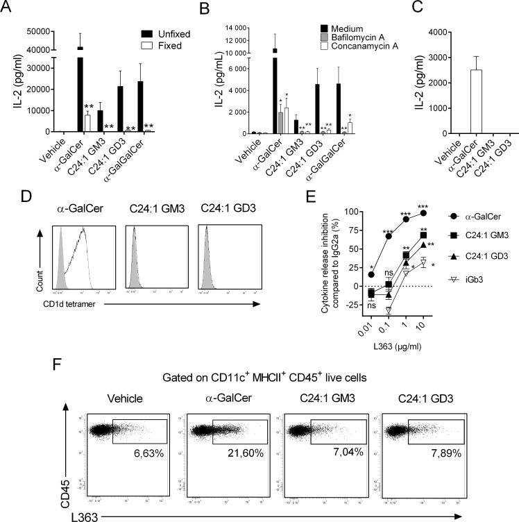
Fig 6. Influence of the DC intracellular pathway on the recognition of gangliosides by the iNKT TCR.
(A) Fixed or unfixed DCs were cultured with the mouse iNKT hybridoma DN32.D3 in presence of purified glycolipids (23 pM for α-GalCer and 6.5 μM for gangliosides) or vehicle for 24 h. (B) Lysosomal acidification inhibitor–pretreated or not DCs were cultured with DN32.D3 in presence of glycolipids or vehicle. (C) Plate-bound CD1d dimers loaded with various glycolipids were cultured with DN32.D3 hybridoma for 24 h. (A–C) iNKT cell activity was evaluated as in Fig 5A. Data represent the means ± SEM of two experiments performed in triplicates. (D) DN32.D3 hybridoma was stained with PE-conjugated CD1d tetramers loaded or not with various glycolipids. Dot plots are representative of one experiment out of two. (E) DCs were cultured with DN32.D3 in presence of purified glycolipids (23 pM for α-GalCer, 1.5 μM for iGb3, and 6.5 μM for gangliosides) or vehicle for 24 h with increasing concentrations of L363 mAb or isotype control. NKT cell activity was evaluated as in Fig 5A. The dose-dependent L363 inhibition normalized to isotype control for each glycolipid is represented. Means ± SEM of two experiments performed in triplicates are shown. (F) Unloaded or glycolipid-loaded DCs were subjected to L363 mAb or Ig-control staining. Dot plots are representative of one experiment out of two for each condition. **P < 0.01; *P < 0.05. Underlying data used in the generation of this figure can be found in S1 Data. α-GalCer, α-galactosylceramide; CD1d, cluster of differentiation 1d; DC, dendritic cell; Ig, immunoglobulin; iGb3, isoglobotrihexosylceramide; iNKT, invariant natural killer T; mAb, monoclonal antibody; NKT, type I natural killer T cell; ns, not significant; PE, phycoerythrin; TCR, T-cell receptor.

