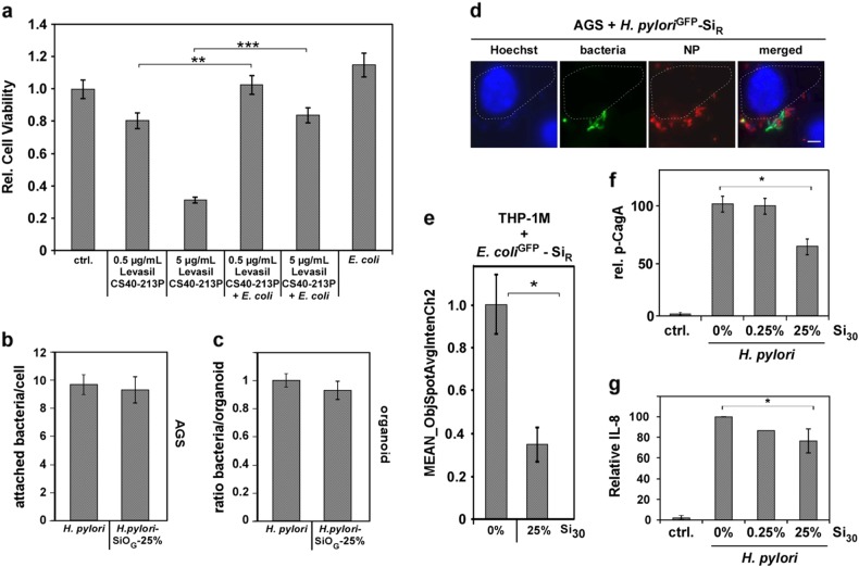
Fig. 3.
NP-binding impacts bacterial pathobiology and fate. a Adsorption of Levasil CS40-213P to bacteria reduced NP toxicity. 2 × 10⁵ human gastric epithelial (AGS) cells were either exposed to 0.5 or 5 µg CS40-213P or to 0.5 or 5 µg CS40-213P pre-incubated with 1 × 10⁷ bacteria for complex formation. Cell vitality was assessed after 6 h. b Live cell fluorescence microscopy visualizes attachment of NP–bacteria complexes. AGS cells were exposed to NP–H. pyloriGFP complexes and analyzed 16 h later. Scale bar 10 µm. c NP-coating does not affect cellular attachment of H. pylori. AGS cells were exposed for 90 min to SiR–bacteria complexes. Attachment was analyzed by confocal fluorescence microscopy. Assays were performed in triplicates, each with a minimum of 100 cells examined. NP–H. pyloriGFP complexes were prepared applying Si30 concentrations estimated to maximally cover approximately 25% of the bacterial surface (1 × 10⁸ bacteria, 600 µg/mL Si30; 10 min PBS). d 3D gastric organoids from normal human corpus mucosa were infected with pristine bacteria or SiR NP–H. pyloriGFP complexes and analyzed by confocal fluorescence microscopy 8 h later. Bacteria and SiR–H. pylori complexes attached to 3D organoid structures equally well. NP–H. pyloriGFP 25% complexes: 1 × 10⁸ bacteria, 600 µg/mL Si30; 10 min PBS. e NP-coating reduces cellular uptake of bacteria into human THP-1M macrophages. Automated microscopy demonstrates reduced internalization of SiR–bacteria complexes. A minimum of 1000 cells was analyzed/well. Complexes 25%: 1 × 10⁸ E. coli, 600 µg/mL Si30, 10 min PBS. f Densitometric quantitation of phosphorylated CagA normalized to β-actin levels in all four experiments. Cells were infected with H. pylori or NP–H. pyloriGFP complexes. At 4 h post infection, CagA and phosphorylated CagA (p-CagA) were analyzed in cell lysates by specific antibodies. g Coating of H. pylori with silica NPs (Si30) results in a NP concentration-dependent decrease in IL-8 secretion. IL-8 was quantified by ELISA in AGS cell supernatants (n = 4). The amount of IL-8 in the sample H. pylori without NPs was set to 100%

