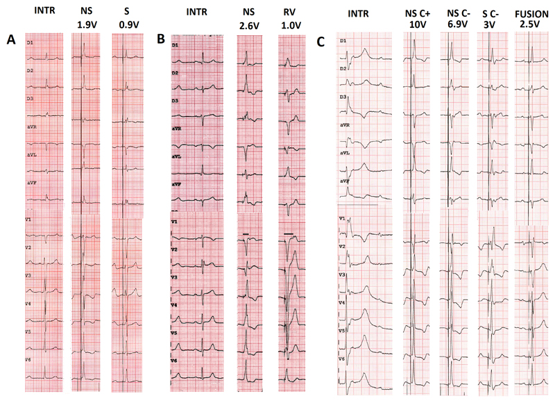
Figure 1.
Examples of types of His capture at different programmed pacing outputs in three patients. A. Patient in atrial fibrillation awaiting atrioventricular nodal ablation. Transition from non-selective to selective His capture. Note the presence of a pseudo-delta wave without an isoelectric interval after the pacing spike with non-selective capture, visible in aVL and the precordial leads B. Patient with exercise-induced Mobitz 2 atrioventricular block and narrow intrinsic QRS. Transition from non-selective His capture to right ventricular capture. Note the increase in stimulus to retrograde P duration with right ventricular capture compared to during His bundle capture (horizontal lines in lead V1). C. Patient in sinus rhythm with complete atrioventricular block and ventricular escape rhythm. Selective capture without correction shows a typical right bundle branch block pattern. Note how it is difficult to distinguish between non-selective capture with and without correction (a difference in QRS morphology is visible in lead II). The patient had a biventricular pacemaker with an atrial, right ventricular, and His lead (connected to the left ventricular port). Due to high capture thresholds for non-selective His capture, fusion pacing with sequential selective His capture (threshold was 1.5V/0.5ms) and right ventricular pacing was programmed.
INTR: intrinsic rhythm; NS: non-selective His capture; S: selective His capture; C+: with correction; C-: without correction.

