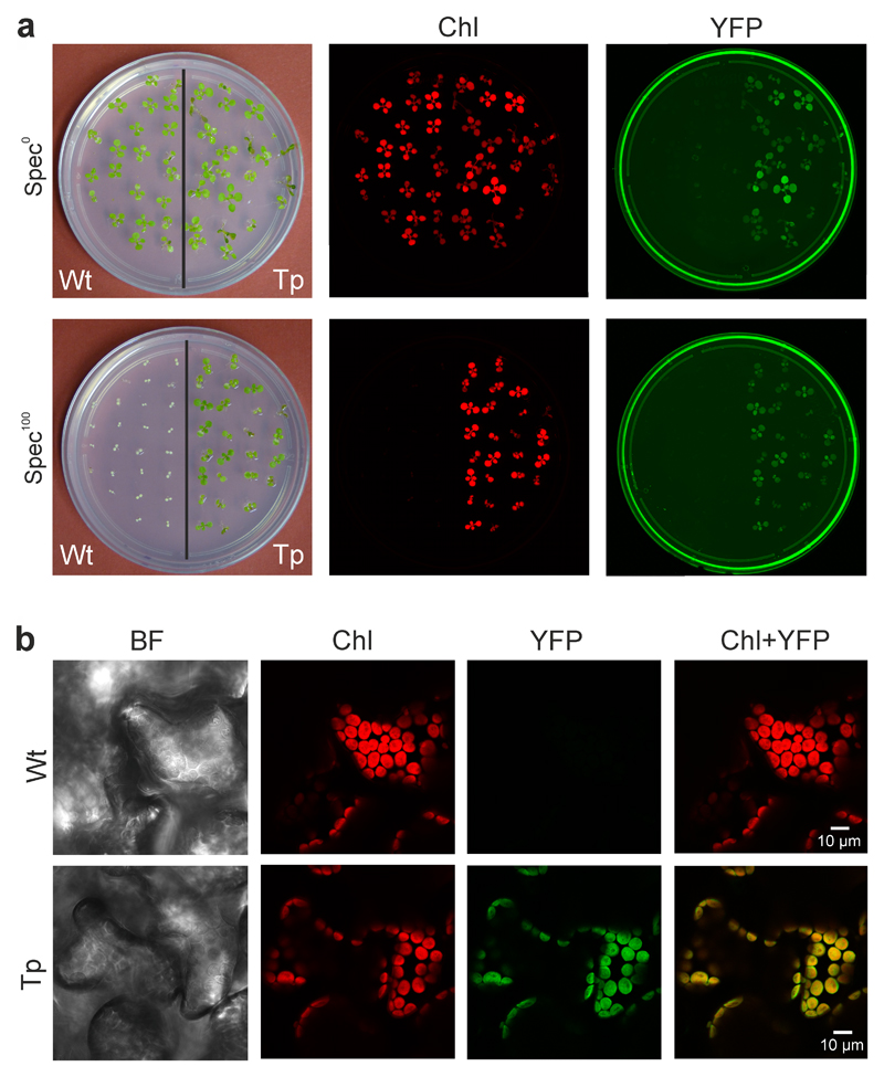
Fig. 4.
Expression of the YFP reporter in transplastomic At-Δa-JF1151 plants. (a) Chlorophyll and YFP fluorescence of wild-type (Wt) and transplastomic (Tp) seedlings grown in the absence (Spec0) or presence (Spec100) of spectinomycin. The plates were scanned with a Typhoon imager that produces a red image of the chlorophyll fluorescence and a green image for the (yellow) YFP fluorescence. The images were taken 20 days after sowing. (b) Confirmation of chloroplast YFP expression in leaf mesophyll cells by confocal laser-scanning microscopy. BF: bright-field image; Chl: chlorophyll fluorescence; YFP: YFP fluorescence (colored in green, to match the color of the Typhoon image); Chl+YFP: overlay of the chlorophyll and YFP fluorescences. These experiments were repeated independently three times with similar results.

