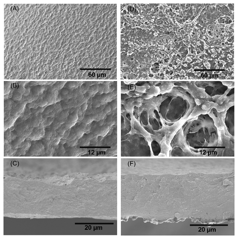
Figure 3.
SEM images of the morphology of the upper side of the [CHI/ALG/CHI/HA]100 at A) lower and B) higher magnifications. The cross-section of the [CHI/ALG/CHI/HA]100 membrane is presented in C). SEM images of the morphology of the upper side of the [CHI/ALG/CHI/HA-DN]100 at D) lower and E) higher magnifications. The cross-section of the [CHI/ALG/CHI/HA-DN]100 membrane is depicted in F).

