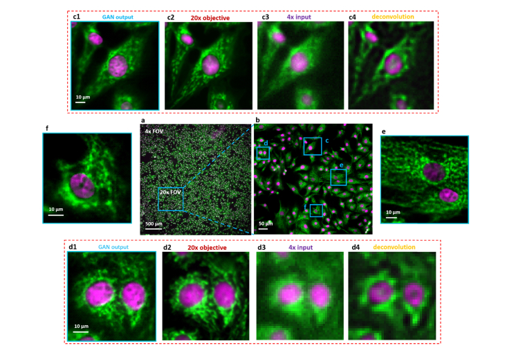
Fig. 6.
Dual-color fluorescence imaging via RFGANM. (a) RFGANM-reconstruction of a wide-FOV fluorescence image of BPAE cells specifically labelled with DAPI and Alexa Fluor-488, at nucleus and skeletons, respectively. (a) and (b) show the imaging FOV of 4 × (which RFGANM inherits) and 20 × objectives (high-resolution conventional microscopy), respectively, by using a sCMOS camera (sensor area 1.33 × 1.33 cm). (c1), (d1), (e) and (f) High-resolution views of the selected regions (blue) in (a). (c2) and (d2) High resolution iamges taken under a conventional wide-field fluorescence microscope with a 20 × /0.45 objective. (c3) and (d3) Low resolution inputs taken under 4 × /0.1 objective. (c4) and (d4) The deconvolution results of (c3) and (d3) respectively.

