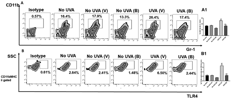
Figure 2. Baicalin decreases CD11b+ Gr1+ myeloid cell and TLR4 expression on CD11b population in UVA irradiated mice.

Mice were sacrificed 24hr post UVA exposure, single cell suspensions of skin (dermal/epidermal) were prepared from dorsal skin. A. Cells were stained with anti-mouse CD11b-APC and anti-mouse Gr-1-PE antibodies. The number of CD11b+Gr1+ cells, was analyzed by flow cytometry. A1. There were significantly less percentage of Gr1+CD11b+ myeloid cells (**p<0.01) in the skin of mice treated with baicalin than in vehicle treated mice. The histograms depict the mean ± SD of cell percentages per group. Mice not exposed to UVA radiation were used as controls (No UVA). B. The expression of TLR4 was also determined on the surface of CD11b+MHCII+ cells using flow cytometry. B1. There were significantly less percentage of TLR4 expression (**p<0.01) in the skin of mice treated with baicalin than in vehicle treated mice. The histograms depict the mean ± SD of cell percentages per group. Mice not exposed to UVA radiation were used as controls (No UVA). N=5; experiment was repeated twice.
