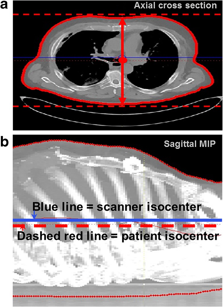
Fig. 2.
3D region growing to extract patient isocenter. a Axial view of region growing to extract patient isocenter for each slice, defined as the midpoint between the highest and lowest points (red dashed lines) of the extracted patient skin contour. b Sagittal MIP is created to demonstrate vertical height measurement of consecutive axial images and demonstrate scanner isocenter does not coincide with patient isocenter (blue line: scanner isocenter, dashed red line: patient isocenter)

