Figure 2.
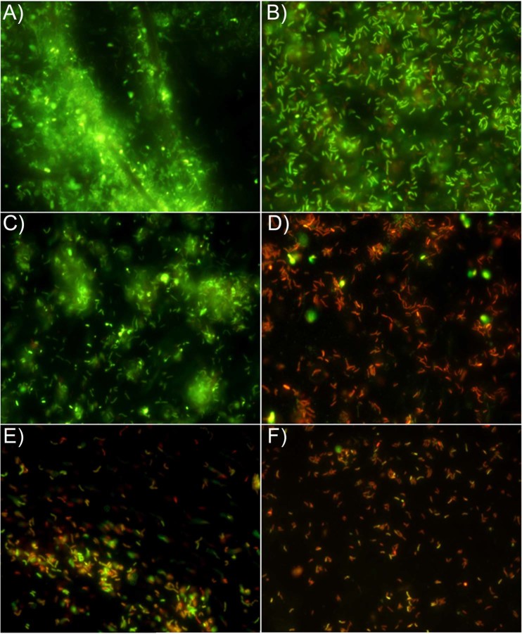
Representative images of Live/Dead staining of H. pylori 11F/11 biofilms. (A,B), control images (viable cells embedded in abundant biofilm were highlighted in A and viable rod shaped cells in the biofilm were highlighted in B). (C), biofilm treated with 1/4 MIC of P. vera L. ORS (prevalent viable coccoid cells in small aggregates). (D), biofilm treated with 1/8 MIC of LVX (prevalent dead rod cells without considerable aggregation). (E,F), biofilm treated with sub-synergistic concentrations of P. vera L. ORS and LVX showing their combined anti-adhesive and killing effect. (E), biofilm treated with 1/8 synergistic concentration of P. vera L. ORS (190 mg/l) and LVX (0.12 mg/l). (F), biofilm treated with 1/16 synergistic concentration of P. vera L. ORS (90 mg/l) and LVX (0.25 mg/l). Sessile population in biofilms stained in red (Propidium iodide) shows a damaged membrane (dead cells), whereas green stained bacteria (SYTO 9) represent viable cells. The images observed at fluorescent Leica 4000 DM microscopy (Leica Microsystems, Milan, Italy) were recorded at an emission wave length of 500 nm for SYTO 9 and of 635 nm for Propidium iodide and more fields of view randomly were examined. Original magnification, 1000X.

