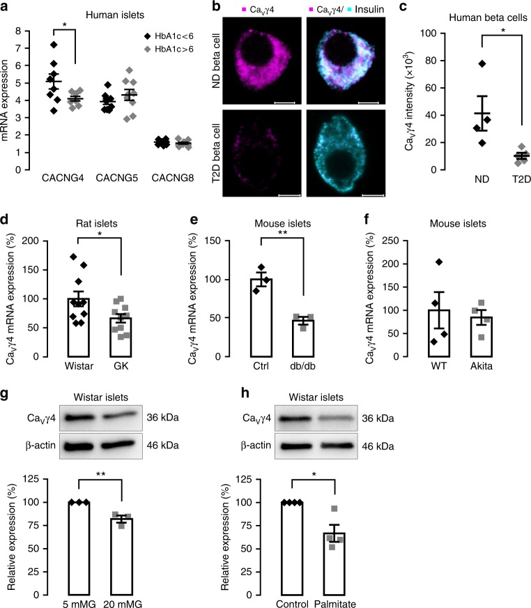
Fig. 1.
Decreased CaVγ4 expression in beta cells in diabetes and in response to glucotoxicity. a CaVγ4 (CACNG4), CaVγ5 (CACNG5), and CaVγ8 (CACNG8) mRNA expression in human islets from donors with HbA1c <6 and >6. n = 8 or 9 donors with age and gender matched, *p = 0.045 (CACNG4), p = 0.294 (CACNG5), p = 0.395 (CACNG8). b Representative immunofluorescence images of CaVγ4 expression in human islet beta cells from non-diabetic (ND) and T2D donor. Scale: 5 μm. CaVγ4 in magenta and insulin in cyan (7–11 beta cells were analyzed per each T2D donor, and 5–7 beta cells for each ND donor). c Calculation of fluorescent intensity for CaVγ4. n = 4 ND and 5 T2D donors (5–11 beta cells were analyzed per donor), *p = 0.029. d Decreased CaVγ4 mRNA expression in Goto-Kakizaki (GK) rat islets. n = 10 rats each, *p = 0.036. e As in d but in db/db mouse islets. n = 3 mice each, **p = 0.006. f CaVγ4 mRNA expression in wild type and Akita mouse islets. n = 4 mice each, p = 0.727. g Decreased CaVγ4 protein expression in Wistar rat islets cultured at 5 or 20 mM glucose (72 h). n = 3, **p = 0.009. h As in g but cultured with 1 mM palmitate (48 h). n = 4, *p = 0.011. Data are presented as mean ± SEM and were analyzed with two-tailed unpaired Student’s t-test. See also Supplementary Table 1 for the details of the human donors utilized for experiments in this study. WT wild type, Ctrl control

