
Fig. 28.
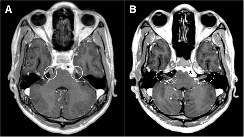
Tuberculosis (TBC). MRI T1-weighted post-gadolinium axial image demonstrates pathologic enhancing tissue involving the basal cistern and ventral surface of the brainstem (asterisks), with leptomeningeal enhancement of bilateral trigeminal (a, circles), facial, and vestibulocochlear nerves (b, dotted circles)