
Fig. 33.
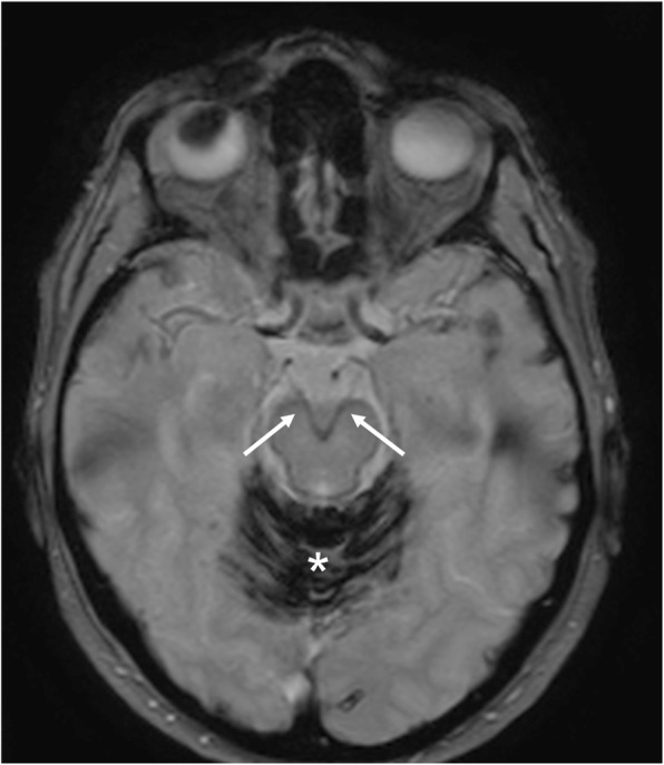
Superficial siderosis. MRI T2*-weighted axial image demonstrates hemosiderin deposition along the course of oculomotor nerves (arrows). Massive hemosiderin deposition is also visible on the cerebellar folia (asterisk)
Official websites use .gov
A
.gov website belongs to an official
government organization in the United States.
Secure .gov websites use HTTPS
A lock (
) or https:// means you've safely
connected to the .gov website. Share sensitive
information only on official, secure websites.
Superficial siderosis. MRI T2*-weighted axial image demonstrates hemosiderin deposition along the course of oculomotor nerves (arrows). Massive hemosiderin deposition is also visible on the cerebellar folia (asterisk)