
Fig. 3.
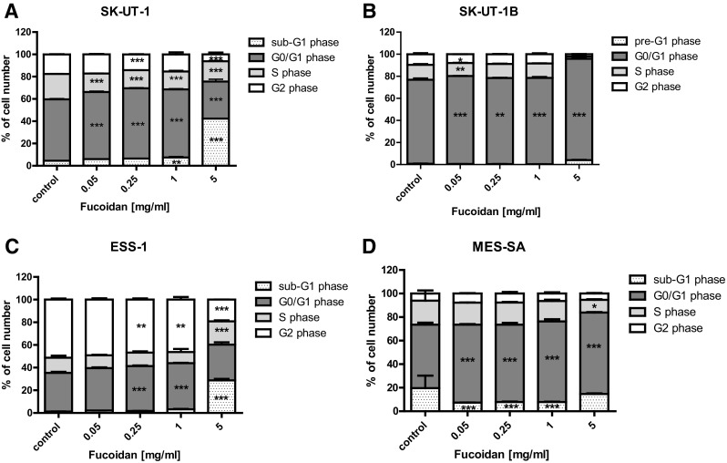
Effect of fucoidan on cell cycle progression in SK-UT-1 (a), SK-UT-1B (b), ESS-1 (c) and MES-SA (d) cell lines. The cell lines were incubated for 48 h with fucoidan (0.05–5 mg/ml) and analyzed by flow cytometry. The results are presented as mean ± SD from three separate experiments. Data were analyzed by flow cytometry and results are expressed as mean ± SD of three separate experiments (n = 6 per each concentration; *p < 0.05, **p < 0.01, ***p < 0.001 versus the control, one-way ANOVA test)