
Figure 3.
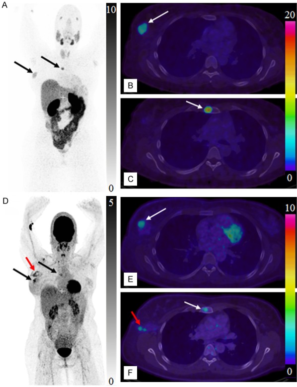
Maximum intensity projection PET images of whole body scan with [68Ga]Ga-ABY-025 (A, 2 h) and [18F]FDG (D). Transaxial PET-CT fused images of the primary tumor (B, E, respectively for [68Ga]Ga-ABY-025 (2 h) and [18F]FDG) and metastasis (C, F, respectively for [68Ga]Ga-ABY-025 (2 h) and [18F]FDG). The black and white arrows indicate known tumor deposits. The red arrow (D, F) indicate post-surgical inflammation after biopsy wherein sentinel lymph node was tumor- and HER2-negative.