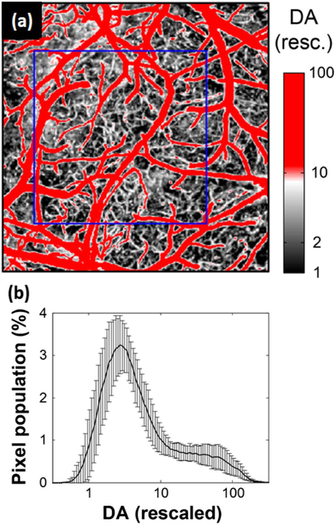
Fig. 3.
Estimation of the capillary perfusion level (CPL) from the rescaled OCT angiogram. (a) DA-based segmentation of capillaries in the rescaled MIP image. The blue box indicated the ROI for estimation of CPL. (b) Histogram of the rescaled DA in log scale (averaged over n = 9 animals). Data are presented as mean ± SD.

