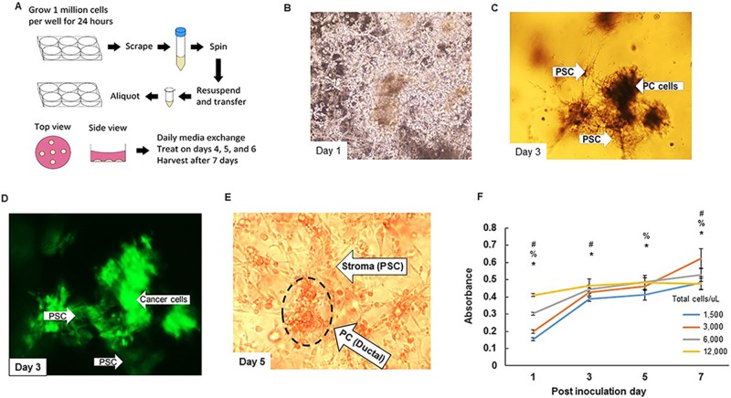
Figure 1. Pictorial representation of cell lines-derived 3D organoid.
A. Scheme of generation of cell lines-derived 3D organoids. Pancreatic cancer and stellate cells (FC1245 and ImPaSC at 1:2 ratio) are grown in monolayer culture for 24 hours at which point we scrape them, centrifuge them, and resuspended them in 1:1 mixture of matrigel: DMEM media supplemented with 10% FBS 1% Pen/Strep and aliquot in 30 μL increments. Cells are treated on PID 4, 5, and 6, and harvested on PID 7 for analyses. B.-D. (B) Microscopic view of organoids on PID 1 with little to no organization present. (C) Microscopic view on PID 3 with significant reorganization present. (D) Immunofluorescence demonstrating congregation of ductal cells. E. Microscopic view of organoids on PID 5 demonstrating the formation of organized structures within the organoids (Supplementary Figure 1, lower magnification, 200x). F. MTT assay demonstrating differential growth for varying cell densities. #p < 0.05 for 3,000 versus 12,000, %p < 0.05 for 3,000 versus 6,000, *p < 0.05 for 3,000 versus 1,500.

