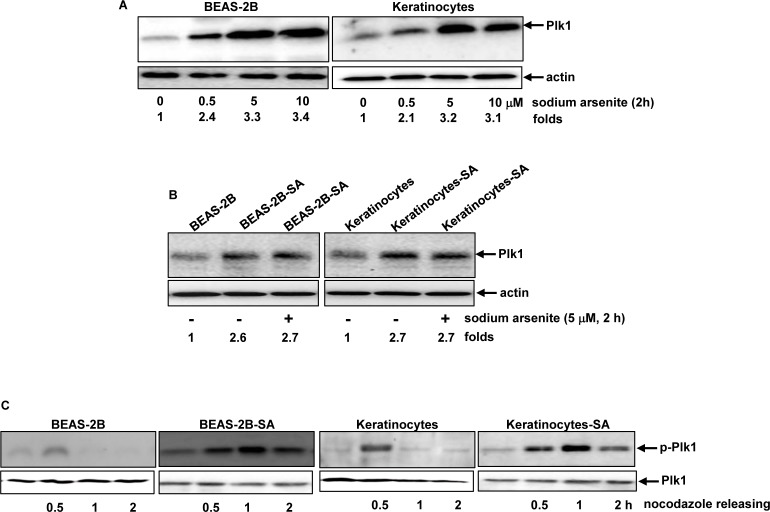
Figure 3. Plk1 activation in response to sodium arsenite treatment.
A. BEAS-2B cells and keratinocytes were treated with different doses of sodium arsenite for 6 h, and Plk expression was examined by immunoblotting analysis. Folds: increases of Plk1 induced by sodium arsenite treatment in comparison with that in untreated controld. B. Cells were stimulated with sodium arsenite for 6 h. Subsequently, the expression of Plk1 was analyzed by immunoblotting. Folds: increases of Plk1 induced by sodium arsenite treatment in comparison with that in untreated controls. C. After released from nocodazole block at different time points, the lysates from the cells were prepared and subjected to immunoblotting for the expression of the phosphorylated Plk1.

