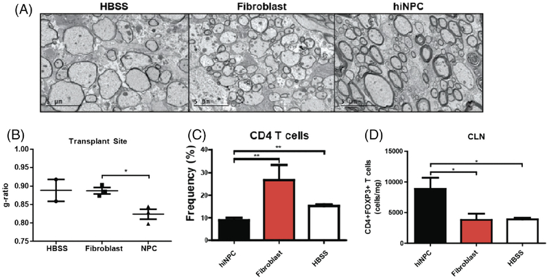
Fig. 3.
Intraspinal transplantation of iPSC-derived NPCs into JHMV-infected mice. A: Focal remyelination in animals transplanted with hiNPCs. Representative electron micrographs of coronal spinal cord sections from HBSS, fibroblast, and hiNPC injected mice. B: Analysis of the ratio of the axon diameter vs. total fiber diameter (g-ratio) confirmed enhanced remyelination. C: Quantification of the percent of CD4+ T cells demonstrated a significant (P < 0.05) decrease in the CLNs of hiNPC transplanted mice compared with controls at 5 days posttransplant (p.t.) D: Quantification of the number of CD4+FoxP3 + Tregs demonstrated a significant (P < 0.05) increase in the CLNs of hiNPC transplanted mice compared with controls at 5 days p.t. Figures derived from Plaisted et al. 2016.

