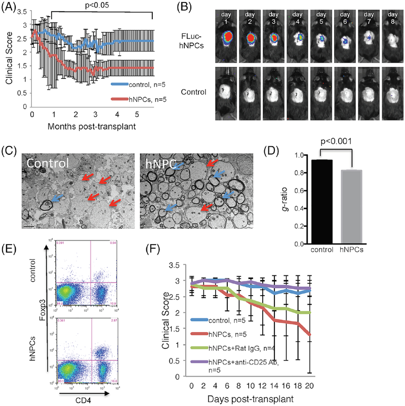
Fig. 4.
Intraspinal transplantation of hNPCs into JHMV-infected mice. (A) Improved (p < 0.05) clinical recovery in hNPC-transplanted JHMV-infected mice was sustained out to 168 days post-transplantation (p.t.) when compared to infected mice treated with vehicle alone. (B) Daily IVIS® imaging of luciferase-labeled hNPCs revealed that following intraspinal transplantation, cells are reduced to below the level of detection by day 8 post-transplantation; representative mice are shown. IVIS® imaging was performed on vehicle-transplanted mice as a control. (C) Representative EM images (1200×) showing increased numbers of remyelinated axons (red arrows) compared to demyelinated axons (blue arrows) in hNPC-transplanted mice compared to control mice. (D) Calculation of g-ratio, as a measurement of structural and functional axonal remyelination, revealed a significantly (p < 0.001) lower g-ratio (indicative of remyelination) in hNPC-treated mice compared to control mice at 3 weeks pt. (E) Quantification of Treg numbers in spinal cords of mice indicated a significant (p < 0.05) increase in numbers of Tregs in hNPC-transplanted mice versus controls between 8–10 days post-transplantation. (F) hNPC-transplanted mice receiving anti-CD25 antibody (purple line) did not display recovery in motor skills as compared to either hNPC-treated mice (red line), hNPC-treated mice receiving isotype-matched control antibody (green line), or vehicle control mice (blue line). Figures derived from Chen et al., 2014.

