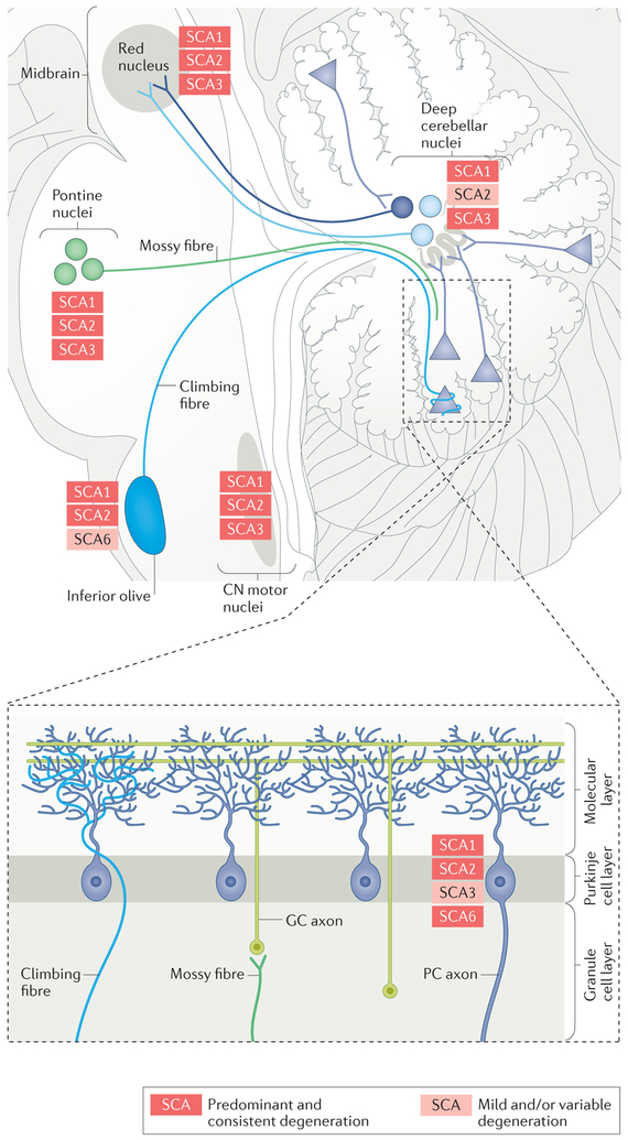
Figure 1 |. Components of the cerebellar circuitry showing degeneration in the polyglutamine spinocerebellar ataxias.
The different spinocerebellar ataxias (SCAs) have similar but not identical patterns of pathological involvement, shown here for SCA1, SCA2, SCA3 and SCA6. The figure shows the areas that are consistently affected in the different SCAs (red), as well as those that are mildly or variably affected in the different SCAs (pink). The areas and cell types in the cerebellar circuitry that are affected include the cerebellar cortex (particularly Purkinje cells (PCs), the cell bodies of which are depicted by triangles here), the inferior olive and its climbing fibres, the pontine nuclei and their mossy fibres, the deep cerebellar nuclei, the red nucleus and the cranial nerve (CN) motor nuclei. GC, cerebellar granule cell.

