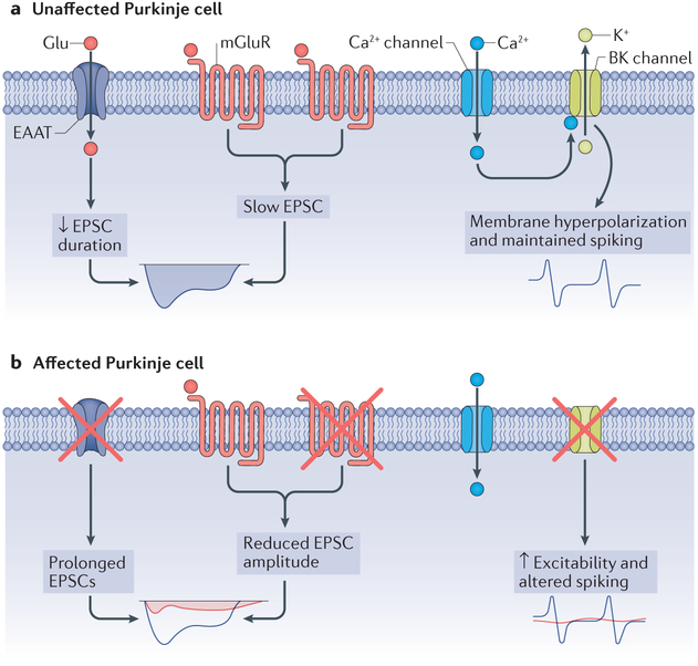
Figure 4 |. Alterations in Purkinje cell electrophysiology in spinocerebellar ataxias.
Concurrent with the motor dysfunction that occurs in the spinocerebellar ataxias (SCAs), and before the onset of substantial cellular morphological alterations, the expression levels and functions of ion channels and receptors are altered in SCA. a | Expression and function of ion channels in an unaffected Purkinje cell. The function of excitatory amino acid transporters (EAATs), which carry glutamate, and metabotropic glutamate receptors (mGluRs) yield slow excitatory postsynaptic currents (EPSCs), whereas normal large-conductance calcium-activated potassium (BK) channel function keeps the cell membrane hyperpolarized, maintaining spiking, b | The reduction in mGluRs and EAATs results in reductions in the amplitude of slow EPSCs (normal currents are shown in blue, and the currents in SCA are shown in red). The reduction in glutamate transporters prolongs the effect of glutamate at the synapse and also prolongs the mGluR-mediated slow EPSCs (red). In addition to alterations in synaptic signalling, the intrinsic excitability of the neuron is altered secondary to a loss of potassium channels. A reduction in BK channel expression and function results in unopposed calcium entry through voltage-gated calcium channels, with impairments in Purkinje neuron spiking (normal spiking indicated in blue; altered spiking indicated in red).

