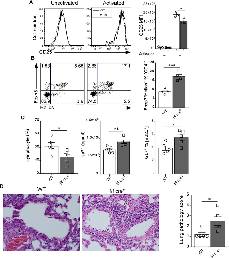
Figure 2. Lack of GARP Affects the Treg differentiation and Decreases the Ability to Control the Inflammation in Pristane-Induced Lupus Model.
(A) Splenic CD4+CD25+ T cells from WT and f/f cre+ were TCR-activated for 72 hr. The expression level of CD25 was determined by flow cytometry. Representative MFI plots and data quantifications are shown (n=3). (B) Flow cytometry analysis of Foxp3 versus Helios expression in splenic CD4+ T cells. Representative dot plots and bar graphs are presented. (C) Female WT (n=5) and f/f cre+ mice (n=5) were given a single i.p. injection of Pristane. Mice were sacrificed 4 months later. Lymphocyte count was determined by complete blood count (CBC), IgG1 serum level was detected by ELISA, and GL7+ % [B220] was measured by flow cytometry. (D) Lungs of WT and f/f cre+ mice were fixed in 4% paraformaldehyde, sectioned, and stained with H&E. Each point represents an individual mouse. Data represent 3–4 independent experiments. Statistical analyses were performed by unpaired Student’s test, *** p<0.001, ** p<0.01, * p<0.05. Error bars represent mean ± SEM.

