Fig. 3.
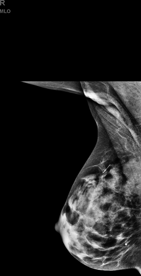
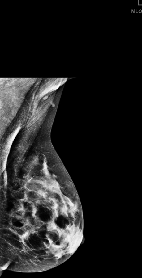
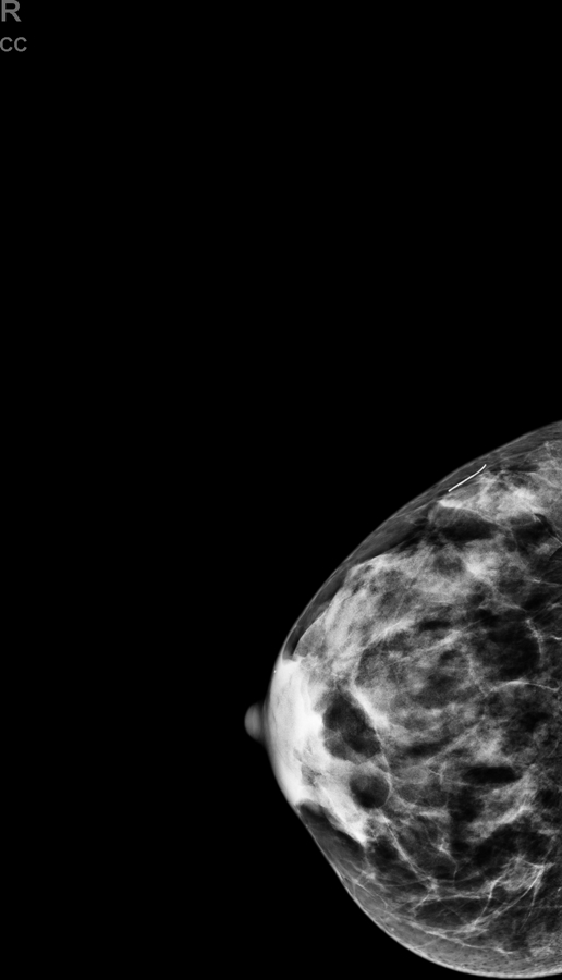
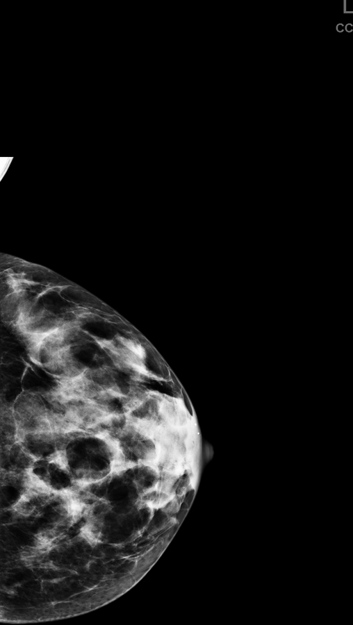
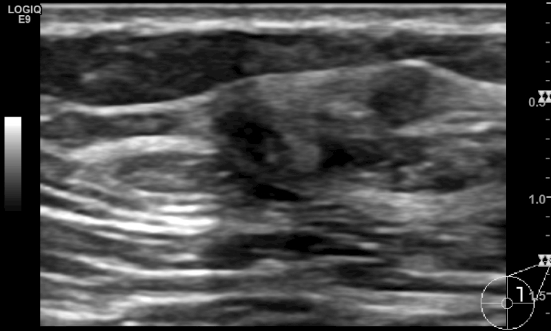
(a,b) Mediolateral oblique and (c,d) craniocaudal mammography in a 52-year-old woman shows dense breast composition type C. (e) Supplemental screening ultrasound shows an irregular hypoechoic 9 mm mass (yellow arrows) in the 2 o’ clock position of the left breast, adjacent to a cyst (green arrow). Biopsy of the irregular mass revealed an invasive, node-negative, intermediate grade, lobular carcinoma (ER positive, PR negative, HER2 negative, Ki-67<1%).
