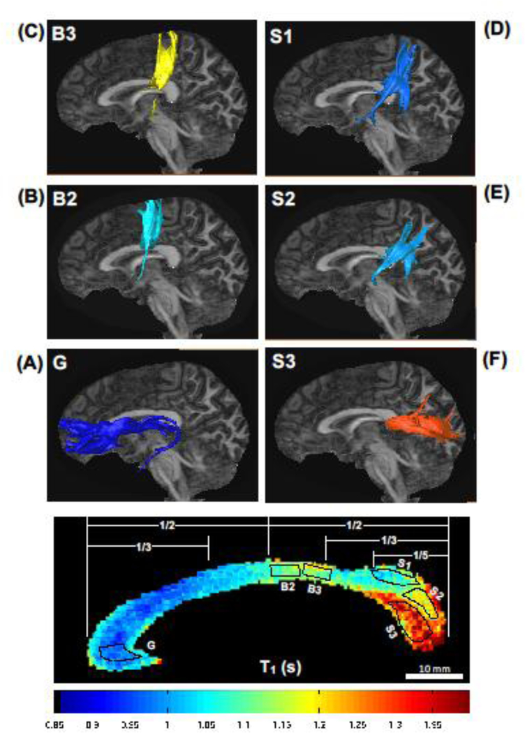
Fig 6.
Diffusion tractography for the human callosal fibers. The callosal fibers from each seeding region of interest (ROI, the dotted areas), which was delineated from the callosal T1 relaxometry map (panel at the bottom), were connected to the structurally projected cortical areas: (A) ROI G to prefrontal lobe; (B) ROI B2 to motor cortex; (C) ROI B3 to sensory cortex; (D-E) ROIs S1 and S2 to parietal-temporal lobes; and (F) ROI S3 to occipital lobe.

