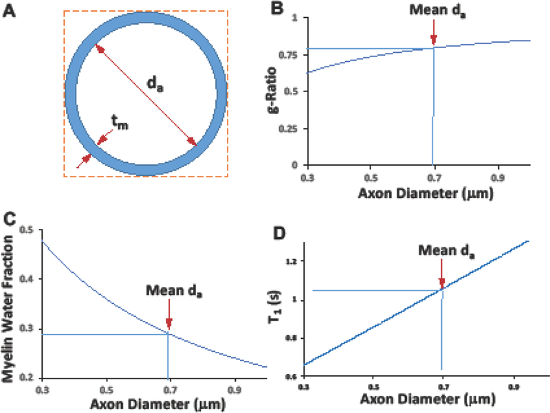
Fig 7.
In vivo model for myelinated axonal diameter. (A) A simplified model of myelinated axon diameter under assumption of an approximation of constant myelin sheath thickness (tm ~ 0.09 m). (B) The estimated microstructure g-ratio , where da is the axon diameter, and its mean value is 0.69 m (pointed by the red arrow) in the human CC (Aboitiz et al. 1992b; Liewald et al. 2014)). (C) The estimated myelin water fraction ( or as a function of axon diameter. (D) The estimated relation between T1 and MWF (T1 = 0.291/MWF +0.05).

