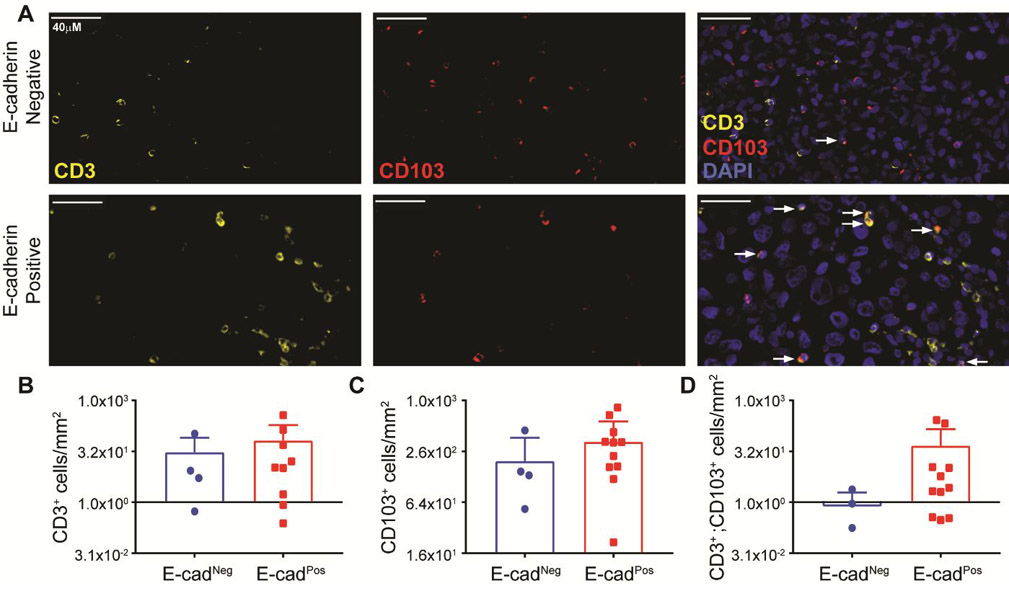
Figure 5. CD103+ lymphocytes infiltrate E-cadherin expressing human metastatic melanoma tumors.

(A) Representative images of immunoflourescent staining against CD3 (yellow, left panel), CD103 (red, middle panel), and overlay with DAPI (blue, right panel). Fifteen tumors were assayed. Four E-cadherin negative (top row) and eleven positive tumors (bottom row) were examined. Tumor E-cadherin status was determined by a dermatopatholgist. E-cadherin negative tumors had no positive tumor cells, while any level of positive staining (low to high) was considered a positive E-cadherin tumor. (B) Number of CD3+ cells per mm2 tumor area. (C) Number of CD103+ cells per mm2 tumor area. (D) Number of CD3+ CD103+ cells per mm2 tumor area.
