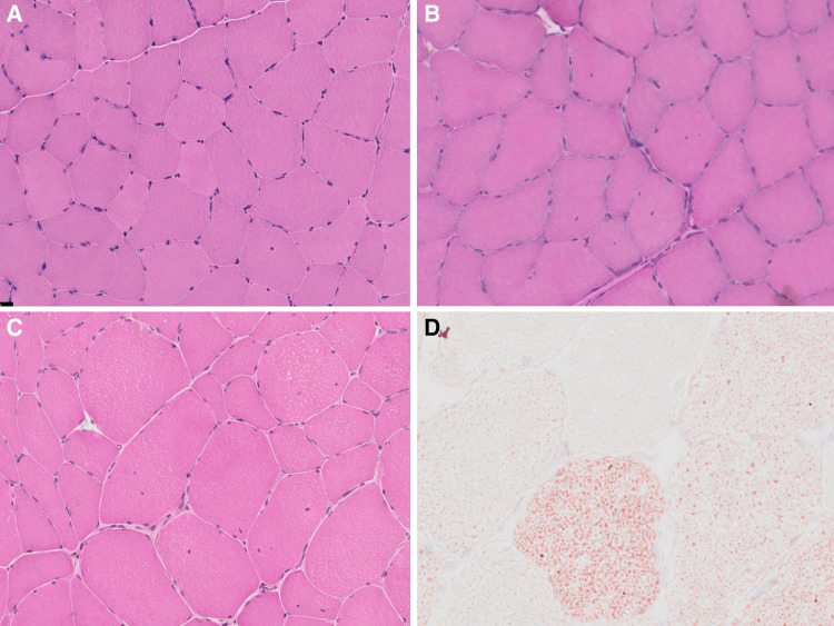
Fig. 1.
Histopathological features—haematoxylin & eosin (H&E) and Oil red O stain: features on H&E (a–c) range from normal appearance (a Patient 7) to increased fibre size variability and an increase in internal nuclei (b Patient 6, and c Patient 4), many of them central (b). On Oil red O stain (d Patient 34), there is marked increase in intracellular lipid, probably related to the timing of the muscle biopsy in relation to the RM episode in the patient

