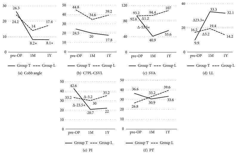
Figure 3.
Mean values of radiological measurements at preoperative, 1 month after the surgery and 1 year of (a) Cobb angle, (b) C7PL-CSVL= C7 plum line, central sacral vertical line, (c) SVA = Sagittal vertical axis, (d) LL = lumbar lordosis, and (e) PI = Pelvic incidence. (f) PT = pelvic tilt. ∗ Difference is significant compared to group L.

