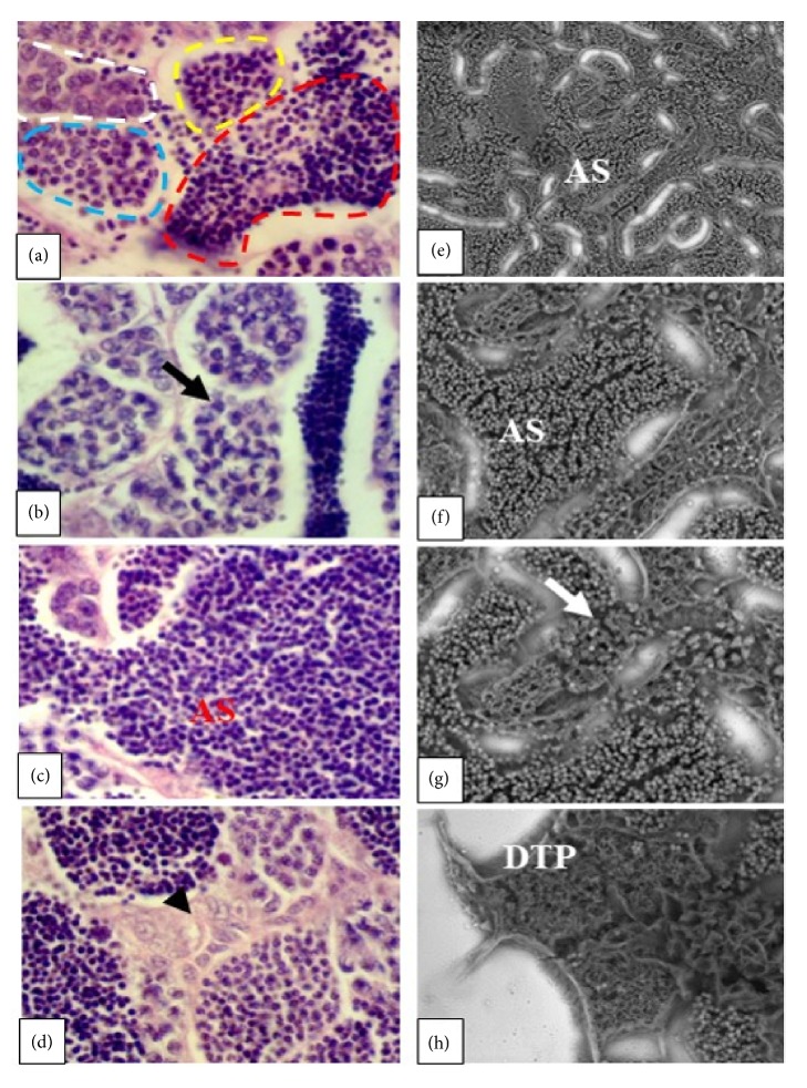
Figure 3.
Longitudinal sections of zebrafish testes. In figures (a) (H&E), (e), and (f) (SEM) normal aspects of zebrafish testes are observed, with mature spermatozoa (red dashed lines), spermatocytes (blue dashed lines), type-II spermatocytes (white dashed lines), and plenty of spermatozoa (AS). In figure (b) (H&E) and (g) (SEM) the development of asynchronous gonads is observed (black and white arrow) in groups treated with EHFAo at 100 and 200 μg/L. In figure (c) (H&E) plenty of mature spermatozoa is observed (groups treated with EHFAo at 100 and 200 μg/L). In (d) (H&E) hyperplasia of interstitial cells is observed (arrowhead), in the group treated with EHFAo at 50 μg/L. In (h) (SEM), testicular parenchyma degeneration is observed (DTP), observed in groups treated with EHFAo at 100 and 200 μg/L.

