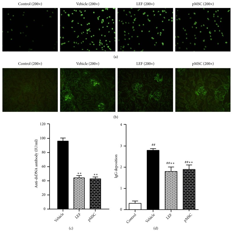
Figure 3.
The serum content of anti-dsDNA antibody and immunofluorescence staining of kidney tissues (fluorescence microscope, 200×). (a) The results of detection by indirect immunofluorescence (IIF) methods; (b) immunofluorescence staining of kidney sections from the vehicle, LEF-treated, and MSCs-treated groups using FITC-conjugated anti-IgG antibodies to compare the effects of pMSC transplantation. (c) The results detected by radioimmunoassay methods. (d) The scores of IgG deposition in the glomeruli are displayed.

