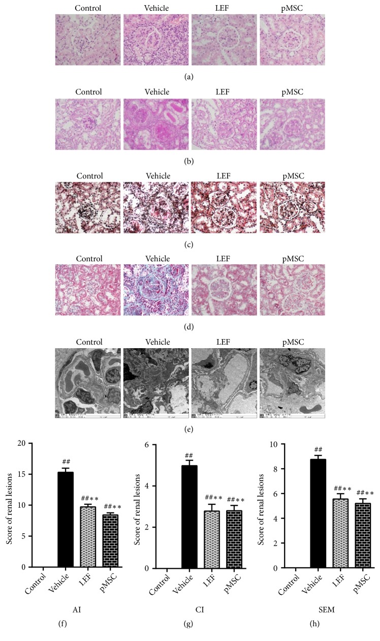
Figure 4.
Histological staining and score of renal tissues. In histological sections of kidneys from an LN-prone murine model with H&E ((a), 400×), PAS ((b), 400×), PASM ((c), 400×), and Masson ((d), 400×) staining, the most common manifestation was intracellular proliferation of capillaries, including mesangial cell and endothelial cell proliferation, with polymorphonuclear neutrophils infiltration often accompanied by the formation of platinum ear-pick or crescent abnormalities. By electron microscopy ((e), 8,000×), electron dense deposits were evident in many parts of the glomerulus, most often in the mesangial area, endothelial cells, and epithelial cells. Occasionally, there were renal tubule or interstitial deposits. The pathological features of the kidney in the two treatment groups were better than those in the vehicle group. (f) The histological scores of the kidneys are shown. Activity index (AI), chronicity index (CI), and score of electron microscopy (SEM).

