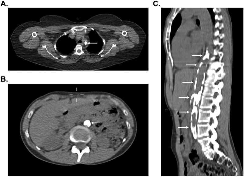
Figure 3.
Representative images of severe vascular calcification in large-vessel vasculitis. Complete occlusion with associated vascular calcification of the proximal left subclavian artery (white arrow) in a patient with Takayasu’s arteritis (Panel A). Near complete occlusion of the abdominal aorta with associated vascular calcification (white arrows) in a patient with Takayasu’s arteritis in axial view (Panel B) and sagittal view (Panel C).

