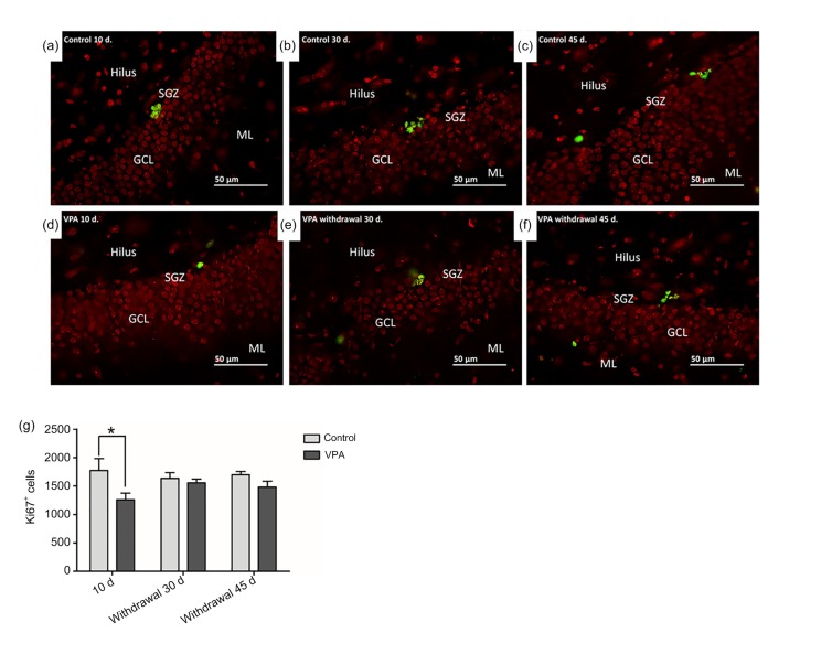
Fig. 3.
Cell proliferation in the sub-granular zone (SGZ) of the dentate gyrus using Ki67-positive staining
(a–f) In the SGZ, the nuclei of Ki67-positive cells were stained green and cells of the dentate gyrus were stained red. (g) Total numbers of Ki67-positive cells (mean±SEM, n=10) in the dentate gyrus were determined from cell counts. The number of Ki67-positive cells in the SGZ of rats receiving VPA was not significantly different from that of rats receiving saline (P>0.05) in both the VPA withdrawal 30 and 45 d groups. However, rats administered with VPA for 10 d showed a significantly lower number of Ki67-positive cells in comparison with the control 10 d group (* P<0.05) (Note: for interpretation of the references to color in this figure legend, the reader is referred to the web version of this article)

