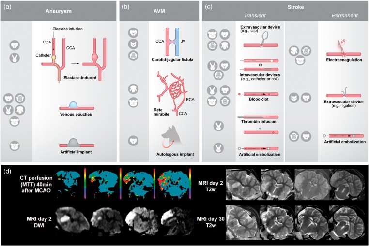
Figure 2.
Illustration of the different techniques for model induction and utilization of clinical imaging procedures. (a) There are three different ways to induce an aneurysm model: elastase-induced aneurysms (aneurysm develops in a closed vessel by elastase infusion), venous pouches aneurysms (preparation of a venous pouch and suturing to artery, usually CCA) and use of artificial implants. Abbreviations: CCA: common carotid artery. (b) AVM models can be created by a carotid-jugular fistula (shunt between CCA and JV), use of RM in pigs and sheep, and use of autologous implants. Abbreviations: JV: jugular vein; ECA: external carotid artery. (c) Ischemic stroke models are based on transient or permanent occlusion of cerebral arteries. Extravascular and intravascular occlusion methods can be discriminated. Extravascular occlusion comprises electrocoagulation and the use of ligation, or occluding devices such as aneurysm clips. Intravascular occlusion can be induced by intravascular devices, such as aneurysm coils, blood clots, thrombin infusion or artificial emboli. (d) A major advantage of large animal models is the compatibility with clinical imaging technologies. Sample images were taken in a sheep after transient MCAO by a surgical clip (3-h occlusion time). Abbreviations: MTT: mean transit time; DWI: diffusion-weighted imaging; T2w: T2-weighted imaging. False color scale indicates MTTS from 0 (purple) to 10 s (red). All images were taken on standard clinical scanner during an in-house study (data not published).

