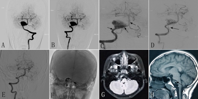
Figure 2.
Images from an 8- to 10-year-old patient with a giant dissecting aneurysm located in the VBJ (case 2). (A,B) Preoperative anteroposterior angiograms of the LVA (A) and RVA (B) showing a giant dissecting aneurysm in the VBJ. (C) Intraprocedural unsubtracted view showing the successful insertion of PEDs (3.5 × 35 mm) (black arrow). (D) Intraprocedural angiogram showing complete occlusion of the LVA by a balloon (black arrow). (E,F) Six-month postoperative angiograms showing the reconstruction of the RVA (E) and stable PEDs (F) (black arrow). (G,H) Six-month postoperative MRI showing a mass effect with a diameter of 20 × 15 mm (black arrow). VBJ, vertebrobasilar junction; LVA, left vertebral artery; RVA, right vertebral artery; PED, pipeline embolization device.

