FIGURE 5.
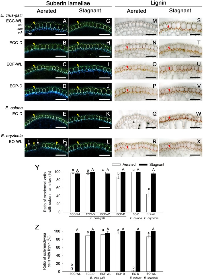
Suberization and lignification in the outer part of roots in Echinochloa accessions under aerated or stagnant conditions. Suberin lamellae and lignin deposits were observed in the basal parts (15–25 mm below root–shoot junction) of adventitious roots of 100–120 mm length. (A–L) Suberin lamellae at the exodermis. Suberin lamellae are indicated as yellow-green fluorescence with Fluorol Yellow 088 (yellow arrowhead). White arrows denote the site of exodermis/hypodermis without suberin lamellae. Blue fluorescence indicates autofluorescence. (M–X) Lignin deposits at the sclerenchyma. Lignin is indicated as orange/red with phloroglucinol-HCl (red arrowhead). CP, cortical parenchyma; epi, epidermis; exo, exodermis; scl, sclerenchyma. Scale bars: 100 μm. (Y) Ratios of cell numbers observed suberin lamellae. (Z) Ratios of cell numbers observed lignin. Means ± SE. n = 4. Different lower-case letters denote significant differences among Echinochloa accessions (P < 0.05, Fisher’s exact test for multiple comparisons). Plants were grown in aerated nutrient solution for 10 days, and then transferred to deoxygenated stagnant 0.1% agar solution or continued in aerated solution for 14 days.

