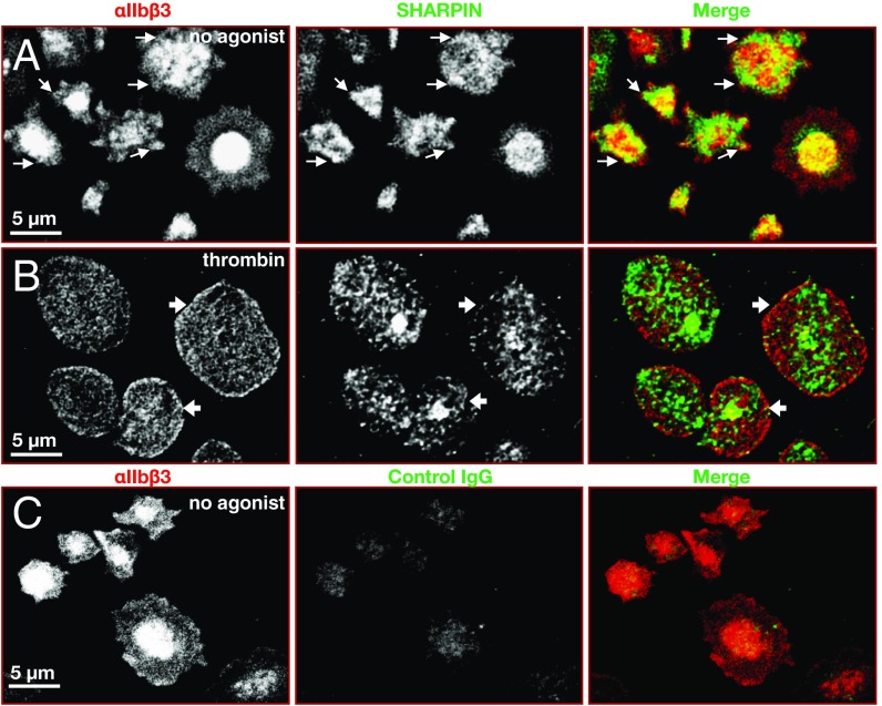
Fig. 2.
Localization of αIIbβ3 and SHARPIN in human platelets. Shown are confocal super-resolution immunofluorescence images of human platelets adherent to fibrinogen, at 100× magnification. Unstimulated (A) or 0.5 U/mL thrombin-stimulated (B) platelets were fixed and stained with antibodies against αIIbβ3, SHARPIN, or (C) control IgG as indicated. Edge localization of SHARPIN and αIIbβ3 was apparent in unstimulated platelets (arrows), but edge localization of SHARPIN appeared to be reduced in thrombin-stimulated platelets (arrowheads). Experiments were conducted three times, with similar results.

