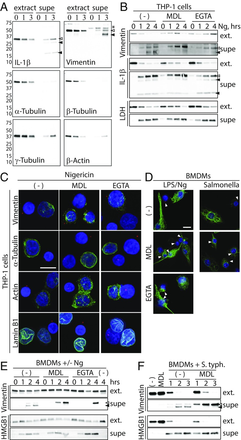
Fig. 5.
Calcium and calpain drive pyroptotic cleavage of vimentin and loss of Intermediate filaments. (A) THP-1 cells were treated with Ng for 0, 1, or 3 h in serum-free medium. Extracts and supernatants were evaluated by immunoblot. Open and closed arrowheads indicate full-length and cleaved proteins, respectively. The asterisk indicates a nonspecific band. Numbers 75–10 indicate molecular weights and position of ladder bands. (B) THP-1 cells were treated for 0, 1, 2, or 4 h with Ng alone or with the calpain inhibitor MDL28170 or EGTA in serum-free medium. Cell extracts (ext.) and supernatants were evaluated by immunoblot. Arrowheads mark inflammasomes. (C) THP-1 cells were treated with Ng alone or with MDL28710 or EGTA. Cells were stained for ASC and with DAPI. Cells were costained for α-Tubulin, actin, lamin B1, or vimentin. Note that vimentin staining in inflammasome-positive cells is enhanced relative to nonpyroptotic cells. (Scale bar: 10 μm.) (D) BMDMs were treated with LPS/Ng and stained for intermediate filaments as in C. Arrowheads mark inflammasomes. (Scale bar: 10 μm.) (E) BMDMs were treated with LPS/Ng and evaluated as in B. (F) BMDMs were treated with S. typhimurium and evaluated as in B.

