Fig. 5.
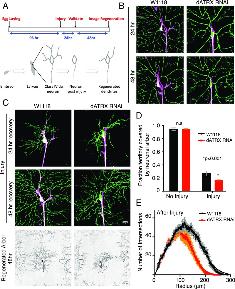
Glial wrapping of somatodendritic surface promotes dendritic regeneration. (A) Schematic representation of the timeline for the balding injury paradigm. (B) Class IV da neuron (green) of RepoGal4 > CD4-tdTomato control larvae and of larvae expressing RepoGal4 > CD4-tdTomato, dATRX RNAi at 24 h and 48 h after no injury. Glia membranes are shown in magenta. (Scale bar, 20 μm.) (C) Dendrites (green) of control and RepoGal4 > CD4-tdTomato, dATRX RNAi expressing larvae at 24 h and 48 h after injury. Glia membranes are shown in magenta. (Scale bar, 20 μm.) The Bottom row shows the entire regenerated arbor of control and RepoGal4 > CD4-tdTomato, dATRX RNAi expressing larvae in grayscale. (Scale bar, 50 μm.) (D) Total coverage of regenerated dendritic arbor in control (black) and RepoGal4 > dATRX RNAi (red) expressing larvae normalized to hemisegment area 48 h after no injury (n = 9, 13 respectively, t test, P > 0.05) and injury paradigm (n = 7 each, t test, P < 0.001). (E) Sholl analysis depicting the number of intersections made by regenerated dendritic branches as a function of radius is shown for the regenerated dendritic arbor 48 h after injury of control RepoGal4 > w1118 (black) and RepoGal4 > dATRX RNAi (red) expressing larvae (n = 7 per condition). The total area under the curve decreased in RepoGal4 > tdTomato, dATRX RNAi larvae compared with control (n = 10 each, 7704 μm2 to 5288 μm2, P < 0.001).

