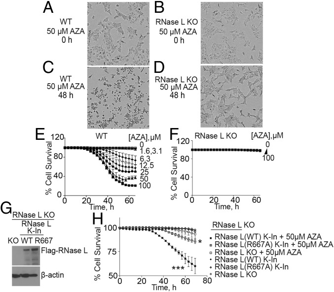
Fig. 1.
RNase L mediates cell death in response to AZA treatment of A549 cells. (A–D) Phase-contrast images of (A and C) WT and (B and D) RNase L KO A549 cells at 0 and 48 h after adding AZA (50 μM) to media. (E and F) Kinetics of cell death of (E) WT and (F) RNase L KO cells after AZA treatment (1.6 to 100 μM). Percent cell survival was determined by real-time imaging with a dual-dye method (SI Appendix, Materials and Methods). Data are the averages ± SD from four identically treated replicates. Three biological replicates were performed, each with a minimum of three technical replicates. (G) RNase L KO cells were transduced with lentiviruses expressing either WT RNase L or a catalytically inactive mutant (R667A) for 72 h to obtain knockin cells. Western blots probed with anti-Flag M2 antibody (Upper), to detect Flag-RNase L, or anti–β-actin antibody (Lower) are shown. (H) Percent cell survival of RNase L KO cells in the absence or presence of lentivirus expressing either WT RNase L or a catalytically inactive mutant (R667A) and incubated without or with 50 μM AZA. Time 0 on the x axis is when AZA was added, 24 h after lentivirus infection. The data are the averages ± SD from three identically treated replicates. Two biological replicates were performed, each with a minimum of three technical replicates. *P < 0.05, ***P < 0.001.

