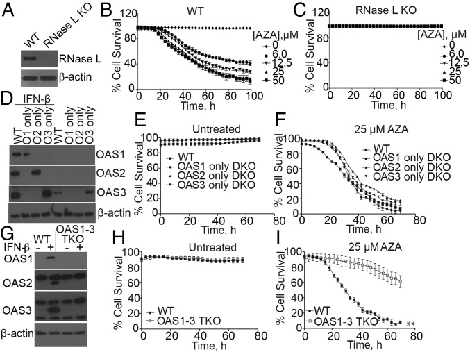
Fig. 2.
Involvement of OAS isozymes and RNase L in the death of HME cells in response to AZA treatment. (A) Western blots of WT and RNase L KO HME cells. (B and C) Percent survival of WT and RNase L KO HME cells, respectively, treated with different doses of AZA. The data are the averages ± SD of four identically treated replicates. Two biological replicates were performed, each with a minimum of three technical replicates. (D) Western blots of OASs from WT and OAS DKO HME cells incubated without or with IFN-β (200 IU/mL for 18 h), using OAS isozyme-specific antibodies. (E and F) Percent survival of WT and DKO cells that were either (E) untreated or (F) treated with 25 μM AZA. The data are the averages ± SD of three identical replicates. Three biological replicates were performed, each with a minimum of three technical replicates. (G) Western blots of WT and OAS1 to 3 TKO HME cells incubated without or with IFN-β (200 IU/mL for 18 h). (H and I) Percent cell survival of WT and TKO HME cells that were either (H) untreated or (I) treated with 25 μM AZA. The data are the averages ± SD of three identically treated technical replicates. Four biological replicates were performed, each with a minimum of three technical replicates. **P < 0.01.

