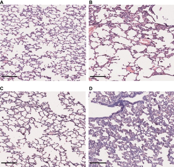
Figure 5.
Lung structure from control and infected hamsters. Microscopic observations of lung sections stained with hematoxylin and eosin obtained from control animals (A) and animals infected with L. interrogans (B), L. mayottensis, (C) or L. borgpetersenii (D) 4 weeks after infection. Scale bars = 150 μm.

