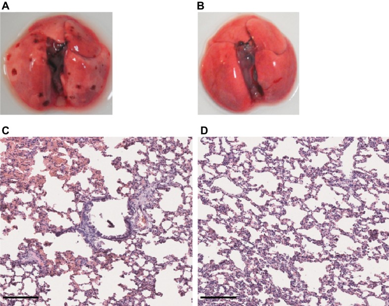
Figure 6.

Lung structure from L. mayottensis-infected hamsters sacrificed at intermediate timepoints. Macroscopic examination of lungs (A,B) and lung sections stained with hematoxylin and eosin (C,D) from L. mayottensis-infected hamsters sacrificed at 10 (A,C) or 17 dpi (B,D). Scale bars = 150 μm.
